Fig. 3.

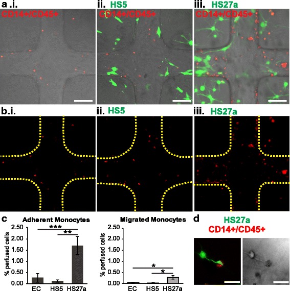
Microenvironment cues change perfused monocyte localization. a Monocytes (red) perfused through unmodified, HS5-, and HS27a-modified vessels adhere to the endothelium (not stained) and transmigrate into the matrix. Scale bars = 100 μm. b Monocytes from a are shown alone along with outlines of vessel walls. Scale bars = 100 μm. c Quantification of monocyte adhesion and migration shows the percentage of cells adhered and migrated within vessels. *p < 0.05, **p < 0.01, *** p < 0.001. d An HS27a cell wraps around a monocyte that has transmigrated into the matrix. EC endothelial cell
