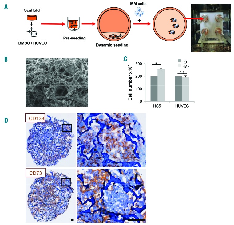
Figure 1.
Generation of a 3D multiple myeloma (MM) microenvironment in bioreactor. (A) Experimental procedure: scaffold is pre-seeded in vitro with bone marrow stromal cells (BMSC)/endothelial cells (HUVEC) and transferred to bioreactor. MM cells are then added and cultured (see Methods section). (B) Scanning electron microscopy analysis of Spongostan (bar=20 mm). (C) Input (t0) (200×103/scaffold) and recovered cell number after 18 hours (h) of 3D culture. Results are mean±Standard Error of Mean of three independent experiments. (D) Immunohistochemistry showing uniform distribution of CD138+ MM cells and CD73+ stroma. Bar=100 mm. *P≤0.05. n.s.: not significant.

