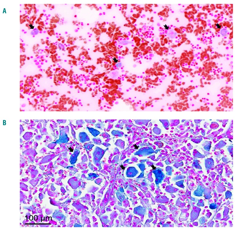
Figure 3.
Iron sequestration in Gaucher cells. Tissue iron content was determined by Perl’s staining of medullar smear (A) and spleen sections (B). The large cells with a laminated aspect were Gaucher cells. The representative images show iron deposition mostly in Gaucher cells. The classical description of Gaucher cells (black arrows) is limited to cells 20–100 μm in diameter with eccentrically placed nuclei and cytoplasm with characteristic crinkles and striations. Images were taken at 20X magnification and a higher magnification (40X).

