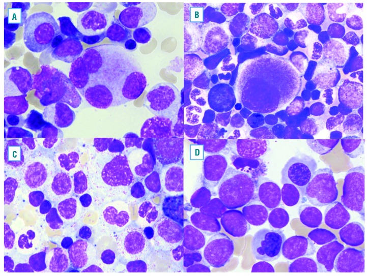
Figure 1.
Examples of typical morphological dysplastic features found in de novo acute myeloid leukemia (AML). Megakaryocytes often show separated lobes (A) and small size micromegakaryocytes (B). Dysplastic changes in myeloid cells, including hypogranular cytoplasm and abnormal nuclear lobation (C). Dysplastic erythroid cells are shown with irregular nuclear contors (D).

