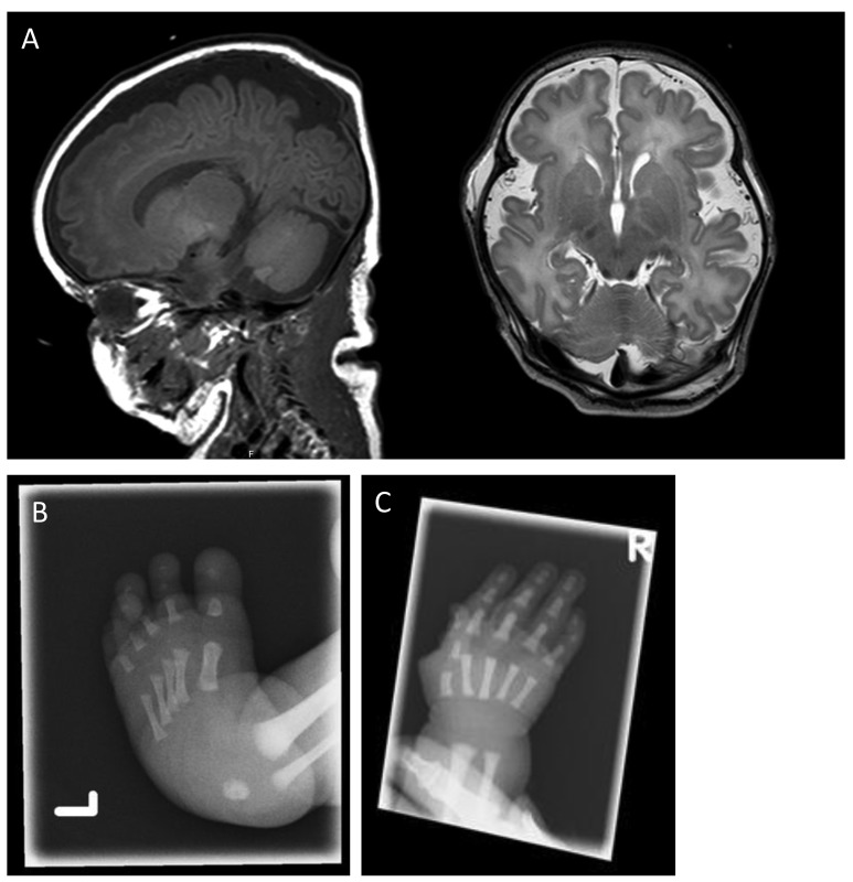
Figure 1.
Cranial MRI and X-ray images of hand and foot of a neonate with syndromic BMF associated with interstitial 3′MECOM deletion. (A) T1-weighted sagittal (left) and T2-weighted axial (right) cranial MRI showing enlarged extracerebral cerebrospinal fluid spaces, and reduced frontal gyrification for the developmental stage (gestational age at birth 38+5 weeks, MRI day 6). (B) X-ray images of left foot showing clubfeet, and (C) a short fifth middle phalanx of the right hand.

