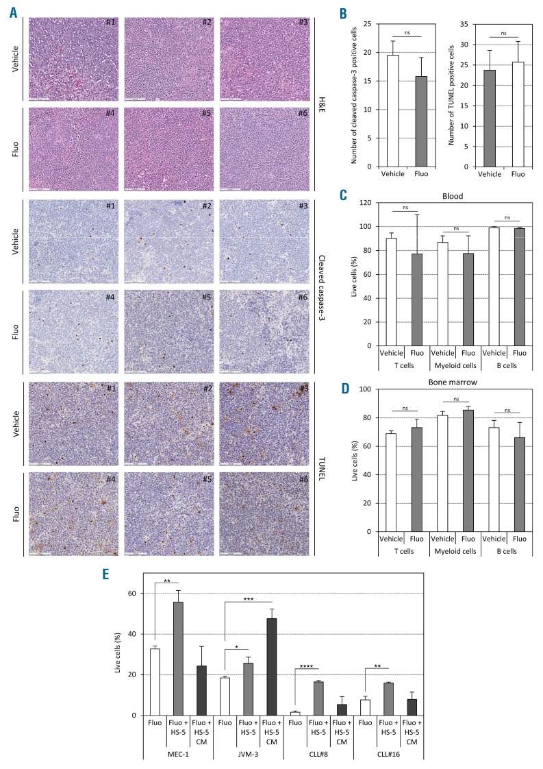
Figure 3.
Fluorizoline (fluo) does not induce apoptosis in vivo. (A–D) AT-TCL1 mice were treated with either 15 mg/kg fluo or equivalent volume of dimethylsulfoxide (DMSO) as vehicle, three times a week for two weeks (5 mice per group). (A and B) Immunohistochemical staining performed on indicated spleen sections using hematoxylin & eosin (H&E), anti-cleaved caspase-3 antibody, and TUNEL assay, respectively (scale bars, 100 μm). The panels show a representative picture for each animal (A) and the respective quantifications (10 fields per mouse in 3 mice per group) (B). (C and D) Cell viability was assessed by annexin-V/7-AAD staining in T cells, myeloid cells and B cells from blood (C) and bone marrow (D) of treated AT-TCL1 mice. (E) Cell viability was assessed in vitro in chronic lymphocytic leukemia (CLL) cells co-cultured on HS-5 stromal cells or with HS-5 conditioned medium only in presence of 5 μM (for cell lines) or 20 μM (for patients’ cells) fluo or equivalent volume of DMSO as vehicle. *P<0.05; **P<0.01; ***P<0.001; ****P<0.0001. ns: not significant.

