
Fig. 3.
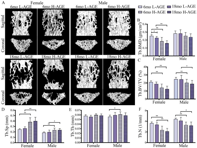
H-AGE intake induced pathological changes in trabecular vertebrae of 6-month-old female mice. (A) 3D μCT images of Tb bone of (top) 6-month-old and (bottom) 18-month-old mice. (B) Tb.BMD, (C) Tb.BV/TV and (D) Tb.Sp demonstrate inferior Tb microstructure in female 6-month-old H-AGE mice. (B-F) Aging dominated diet effects in both sexes. *p < 0.05, **p < 0.001. Tb = trabecular; Tb.BMD = trabecular bone mineral density; Tb.BV/TV = trabecular bone volume fraction; Tb.Sp = trabecular separation; Tb.Th = trabecular thickness; Tb.N = trabecular number; mo = months.