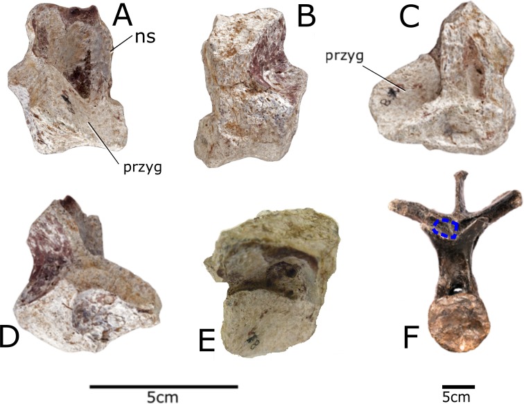
Figure 4. Vertebra of Paranthodon africanus NHMUK R47338.
(A) Anterior; (B) posterior; (C) left lateral; (D) right lateral; (E) dorsal; (F) comparison with dorsal vertebra five of NHMUK R36730 showing location of fragmentary vertebra of Paranthodon. ns, neural spine; przyg, prezygapophysis. Scale bar on left is for (A), (B), (C), (D), and (E). Scale bar on right applies to (F) only. Images copyright The Natural History Museum.

