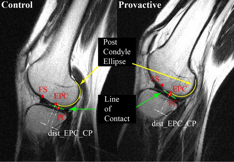
Fig. 7.
Magnetic resonance images of the same knee in the control and provocative positions, showing the tibiofemoral joint contact (green), the elliptical outline of the posterior femoral condyle (EPC) (yellow), the distance from the midpoint of the tibiofemoral line of contact (PC) to the point at which the elliptical outline of the posterior femoral condyle diverges from the cortical bone (dist_EPC_CP) (white); and the femoral sulcus (FS) location. (Reprinted, with modification, from: Boden BP, Breit I, Sheehan FT. Tibiofemoral alignment: contributing factors to noncontact anterior cruciate ligament injury. J Bone Joint Surg Am. 2009 Oct;91[10]:2381-9.)

