Figure 3.
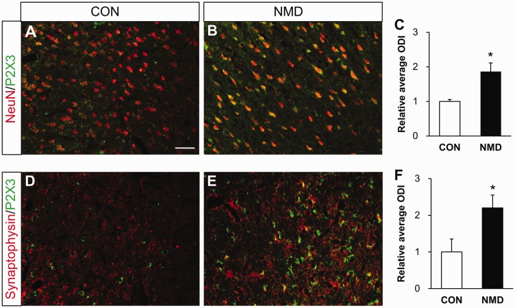
Enhanced P2X3Rs expression in neurons and presynaptic terminals of right IC of NMD rats. (a) Merged immunofluorescence images showed minor co-localization of NeuN (red) and P2X3Rs (green) in right IC of CON rats. (b) Merged images showed many co-localizations of NeuN (red) and P2X3Rs (green) in right IC of NMD rats. (c) Statistic chart indicated that P2X3Rs positive neurons were significantly increased in right IC of NMD rats compared with CONs (n=4 for each group, *p<0.05 vs. CON, two sample t test). (d) Immunofluorescence images showed minor co-localization of synaptophysin (red) and P2X3Rs (green) in right IC of CON rats. (e) Images showed many co-localizations of synaptophysin (red) and P2X3Rs (green) in right IC of NMD rats. (f) Statistic chart showed that P2X3Rs positive synaptophysin was markedly increased in right IC of NMD rats compared with CONs (n=4 for each group, *p<0.05 vs. CON, two sample t test). Bar=100 μm for all photos. CON: control; NMD: neonatal maternal deprivation.

