Figure 4.
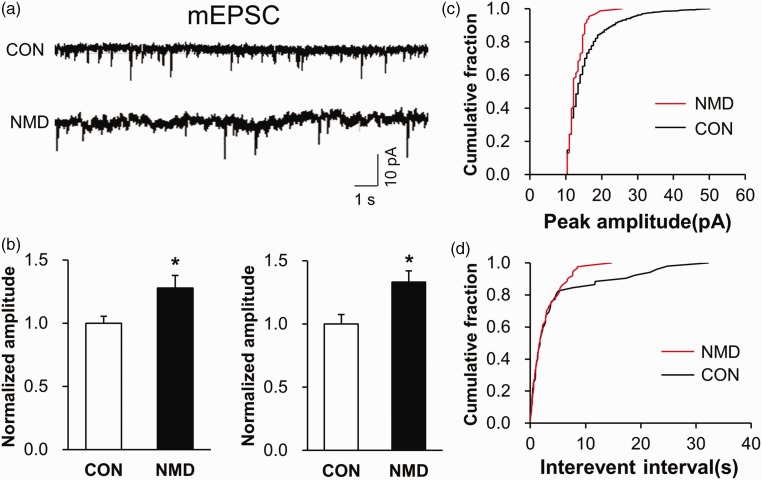
Potentiation of mEPSC in right IC of NMD rats. (a) Recordings illustrating mEPSC of typical neurons in right IC of CON (top) and NMD (bottom) rats. (b) The frequency and amplitude of mEPSC were both greatly increased in right IC of NMD rats compared with CON ones (n=6 cells for CON group, n=8 cells for NMD group, *p<0.05 vs. CON, two sample t test). (c) Cumulative fraction of peak amplitude of mEPSC in an IC pyramidal neuron of NMD and CON rat, respectively. (d) Cumulative fraction of interevent interval of mEPSC in an IC pyramidal neuron of NMD and CON rat, respectively. mEPSC: miniature excitatory postsynaptic current; CON: control; NMD: neonatal maternal deprivation.

