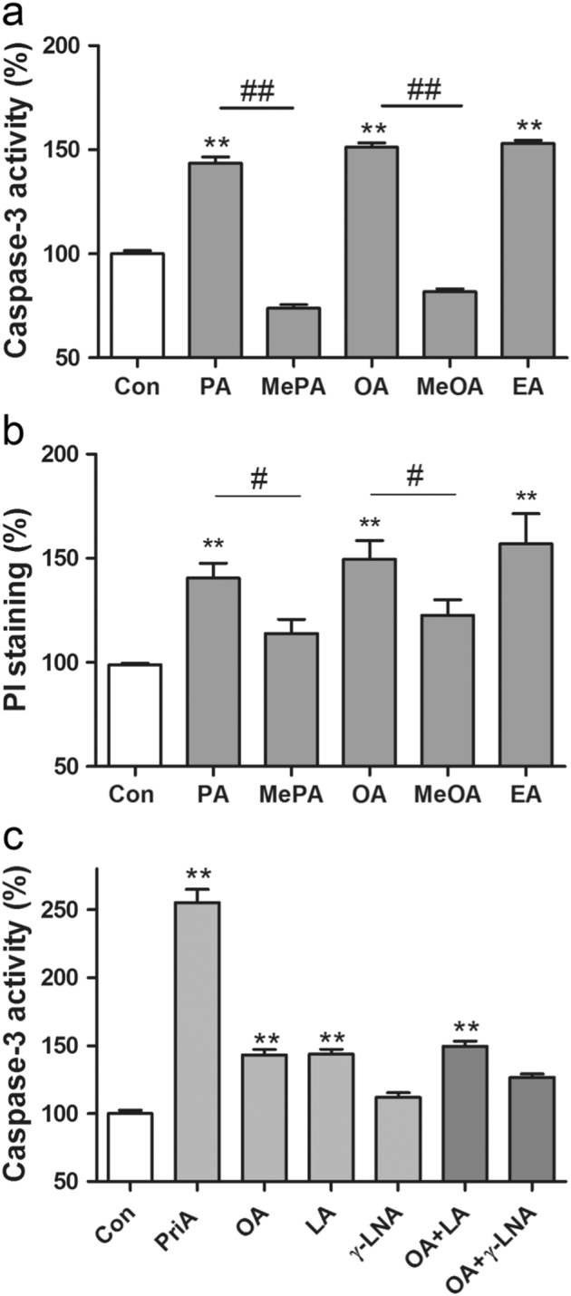
Fig. 2.

Toxicities of PA, MePA, OA, MeOA and EA (trans-OA) a-b and toxicities of the PUFAs LA (C18:2) and γ-LNA (C18:3) in comparison with the MUFA OA (C18:1) and to PriA in human EndoC-βH1 beta cells and their ability to antagonise the toxic effect of OA (C18:1) c. EndoC-βH1 beta cells were incubated for 2 days with PA, MePA, OA, MeOA and EA (trans-OA)(all FFAs 500 µM). Thereafter, caspase-3 activity a and propidium iodide fluorescence b were measured. In addition, EndoC-βH1 beta cells were incubated for 2 days with the polyunsaturated fatty acids (PUFAs) LA (500 µM), γ-LNA (500 µM), and for comparison with the monounsaturated OA (500 µM), as well as the branched-chain PriA (200 µM). Furthermore, combinations of OA with the PUFAs (500 µM each) were incubated. Thereafter, caspase-3 activity was measured c. Data are means ± SEM of 5–8 independent experiments. **p < 0.01 compared with untreated control cells; # p < 0.05, ## p < 0.01 compared with the corresponding non-methylated fatty acid.
