Figure 8.
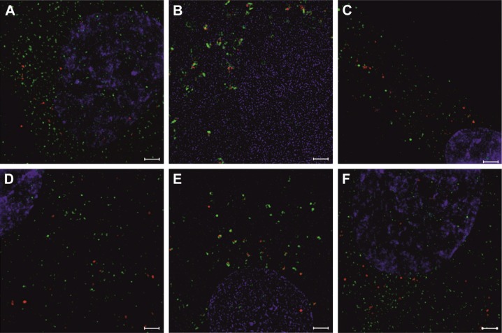
The SR-SIM analysis of hBM-MSCs, 24 hours after their co-culture with EVs previously stained with different dyes.
Notes: EVs labeled with PKH26 (A–C) or tagged with Molday ION (D–F) (red) taken up by hBM-MSCs are visible inside the cells. Coexpression of tetraspanins: CD9 (A and D), CD63 (B and E), and CD81 (C and F) (green) were demonstrated. Cell nuclei were stained with Hoechst (blue). Scale bar =20 μm.
Abbreviations: EVs, extracellular vesicles; hBM-MSCs, human bone marrow mesenchymal stem cells; SR-SIM, super-resolution structured illumination microscopy.

