
Figure 3. Proposed mechanisms for facilitation.
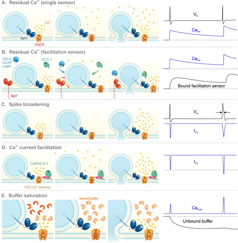
Illustration of the proposed mechanisms for facilitation. Voltage-gated Ca2+ channels (VGCC) open during the first action potential and allow Ca2+ influx (ICa). During the first action potential (AP) Ca2+ influx fails to fully activate Syt1 and trigger fusion.
A) The residual Ca2+ hypothesis: Cares from the first AP adds to Calocal during the second AP. Because the fusion sensor binds Ca2+ in a supralinear manner, a small increase in the Ca2+ signal drives a large increase in p.
B) Residual Ca2+ binds to a facilitation sensor: A high-affinity sensor, distinct from Syt1, binds to Cares in between APs and raises p by interacting with the fusion machinery.
C) Spike broadening: Inactivation of K+ channels during the first AP broadens the waveform of the second AP, prolonging the time-course and total amount of ICa.
D) Calcium current facilitation: Ca2+ from the first AP binds to NCS-1 or CaM, which interact with the IQ-like motif (IM) on the intracellular C-terminus of P/Q channels to increase ICa during the second AP.
E) Buffer saturation: During the first AP there is enough unbound Ca2+ buffer to capture incoming ions and decrease the Ca2+ signal sensed by Syt1 (Calocal). During the second AP the buffer becomes saturated and cannot constrain Calocal.