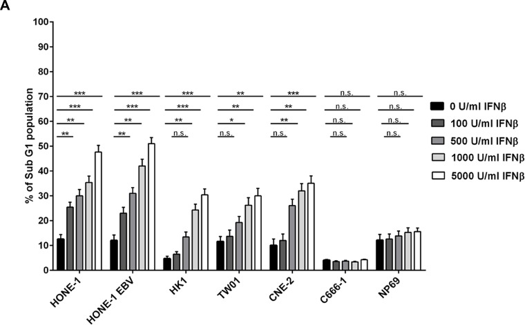
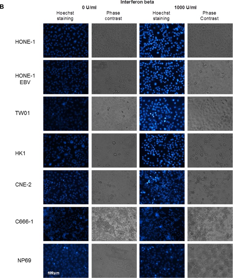
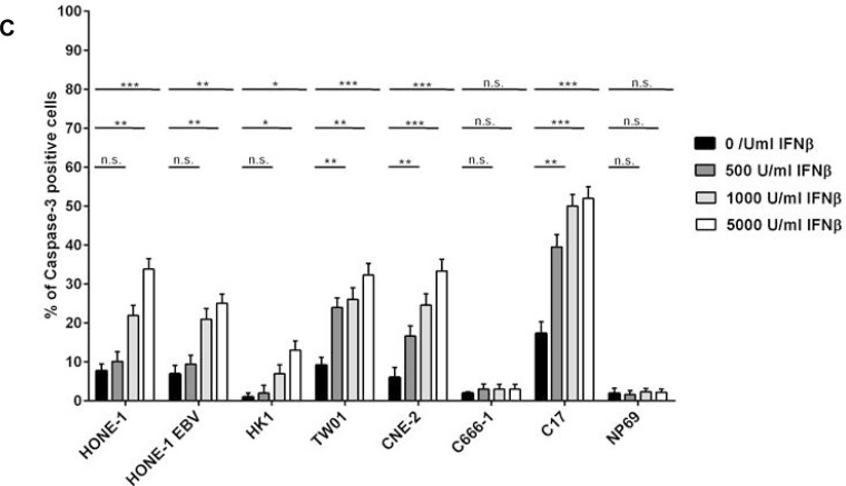
Figure 2. IFNβ induces apoptosis in NPC cells.
(A) Cell cycle analysis. IFNβ induces apoptosis measured by an increase in subG1 in NPC cell lines HONE-1, HONE-1 EBV, CNE-2, HK-1 and TW01. No effect is seen in NPC cell line C666-1 and the nasoepithelial cell line NP69. The data represent the means of three independent experiments and the corresponding standard error. (B) Hoechst 33258 staining. Treatment with IFNβ leads to morphological signs of apoptosis (condensed and fragmented nuclei) in NPC cell lines HONE-1, HONE-1 EBV, CNE-2, HK-1 and TW01, but not in cell line C666-1 and the immortalized nasoepithelial cell line NP69. Morphologic changes were examined under a fluorescence microscope at 200× magnification. Phase contrast images are shown to compare for cell density. (C) Treatment with IFNβ increases the number of cells with activated caspase-3 in the five IFN-sensitive NPC-cell lines and in C17-PDX cells but not in cell line C666-1 and nasoepithelial cells. Quantitative data are reported as means ± S.E.M. (triplicate samples). Data of all experiments are shown at 72 h after incubation with IFNβ. *p < 0.05; **p < 0.01; ***p < 0.001.


