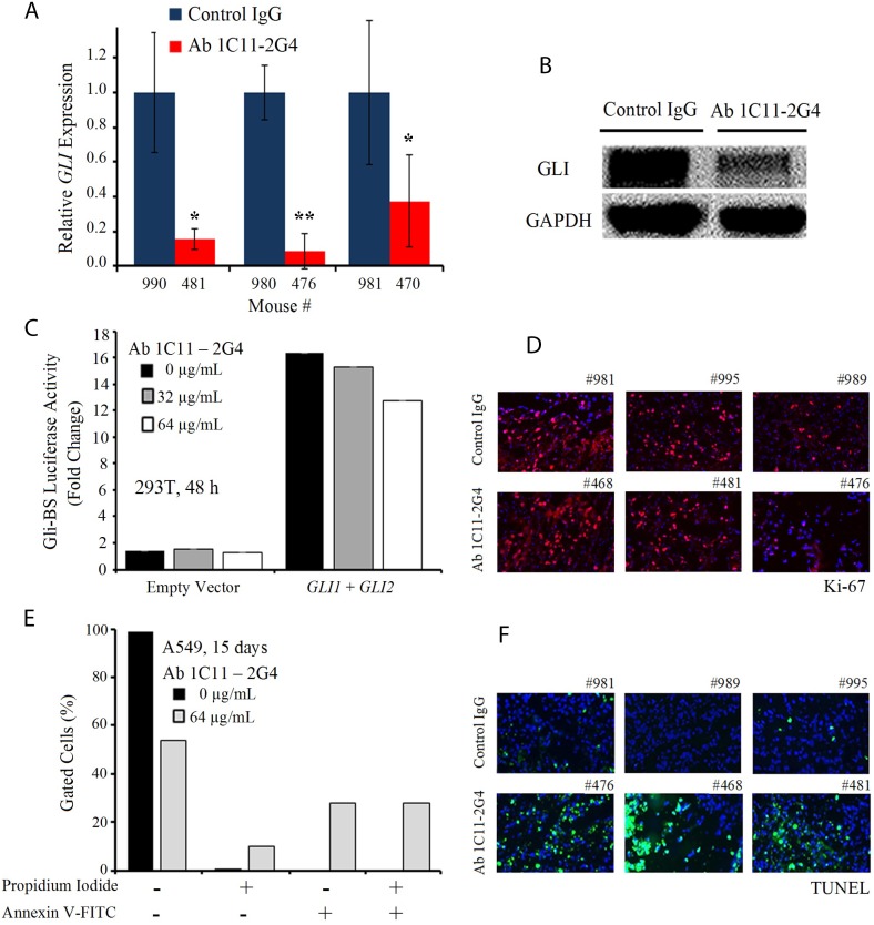
Figure 6. C-term Shh Ab 1C11–2G4 treatment down-regulates Gli and induces apoptosis.
(A) Ex vivo quantitative RT-PCR analysis of GLI levels in A549 harvested tumors treated with IgG controls OR 8 mg/kg Ab 1C11-2G4, 3× per week for 3 weeks. Real-time PCR reactions were performed in triplicate and the data presented as fold change in target gene expression (mean ± SD) after normalization with GAPDH. The results shown are representative of two independent experiments. (B) Corresponding tumors after control IgG OR Ab 1C11-2G4 treatments were lysed and analyzed by Western blot for the expression of GLI. GAPDH was used as a loading control. (C) Empty vector OR GLI1 and GLI2-induced transcriptional activation in 293T cells left untreated or treated with 2 doses of Ab 1C11-2G4 for 48 hours. An expression construct linking the 8 repeats of Gli-binding sites (Gli BS) to a luciferase reporter was used as a surrogate measurement of the Gli-dependent transcription. All measured luciferase activities were normalized to pRL-TK vector activity. The data represent means ± S.D. (D) Control IgG- or Ab 1C11-2G4-treated tumor sections stained for a common proliferation marker, Ki-67. Representative images captured at 20× are presented. (E) A549 cells were treated with 64 μg/mL of Ab 1C11-2G4 for 15 days, stained with Annexin V-FITC/PI and analyzed for apoptosis by flow cytometry. The resulting percentages are presented as a graph. (F) Control IgG- or Ab 1C11-2G4-treated tumor sections stained for apoptosis induction via a TUNEL assay. Representative images captured at 20× are presented.

