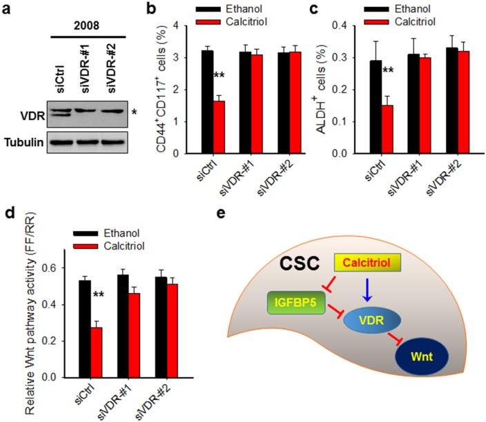
Figure 5. Calcitriol depletes CSCs in the ovarian cancer cell population through VDR-mediated inhibition of the Wnt pathway.
(a) VDR expression is successfully downregulated by VDR siRNA. 2008 cells were transfected with control siRNA or two different VDR siRNA for 2 days. Immunoblotting was conducted to analyze the protein level of VDR. Tubulin was detected as a loading control. *: non-specific band. (b, c) Knockdown of VDR compromises calcitriol-induced depletion of CSCs. 2008 cells were transfected with either control or two different VDR siRNA for 24 h, treated with 5 nM calcitriol for 5 days. FACS was conducted to analyze the percentage of CD44+CD117+ cells (b) and ALDH+ cells (c). N = 3, Bar: SD, **: P < 0.01. (d) Knockdown of VDR blocks calcitriol-induced inhibition of Wnt pathway activity. 2008 cells were treated same as above described, luciferase assay was conducted to determine the Wnt pathway activity. (e) Schematic diagram of the mechanism of calcitriol-induced depletion of CSCs. In CSCs, calcitriol treatment inhibits the expression of IGFBP5, which is an antagonist of the VDR pathway signaling. The reduced expression of IGFBP5 can promote calcitriol-induced activation of the VDR pathway signaling, which further inhibits the Wnt pathway, leading to the loss of stem cells’ properties of CSCs.

