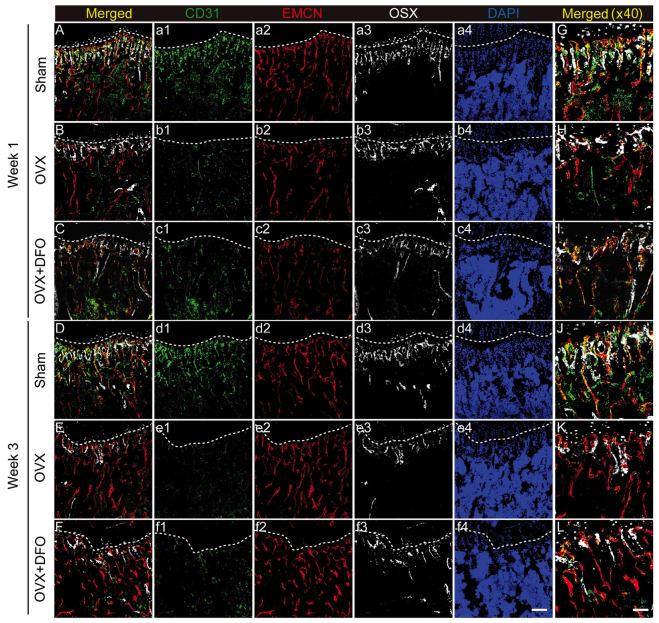
Figure 4.
Changes of type H vessels in osteoporotic mice. (A-F) Tibia immunostaining (×20) for CD31 (green), endomucin (red), osterix (gray) and DAPI (blue) of the sham (A and D), OVX (B and E) and OVX + DFO (C and F) group mice at week 1 (A-C) and week 3 (D-F), respectively. (G-L) Higher magnification (×40) view of metaphysis region. The line indicates the boundary of the metaphysis and growth plate. OVX, ovariectomy; DFO, desferrioxamine. Scale bar, 100 µm in F (f4); 200 µm in L.

