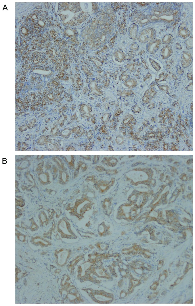
Figure 5.

Representative immunohistochemical staining for TBX2 in prostate cancer. TBX2 was located in the cytoplasm of prostate cancer cells. The expression rates of TBX2 were 75.47% (40/53) in prostate cancer tissue, while 41.51% in tumor adjacent tissue (P<0.01). (A) Positive staining ‘++’ for TBX2 in prostate cancer with well differentiation (×100). (B) Positive staining ‘+++’ for TBX2 in prostate cancer with poor differentiation (×100).
