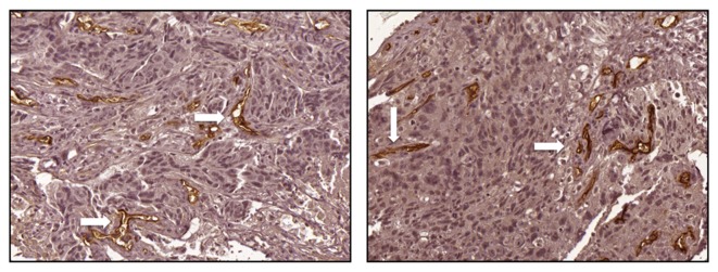
Figure 2.
Representative immunohistochemistry images of increased expression of endoglin in breast tumor samples following neoadjuvant chemotherapy from patients (magnification, ×200). Positive endoglin staining (white arrows) was observed as thin, linear deposits in the membrane and cytoplasm of endothelial cells within the microvessels.

