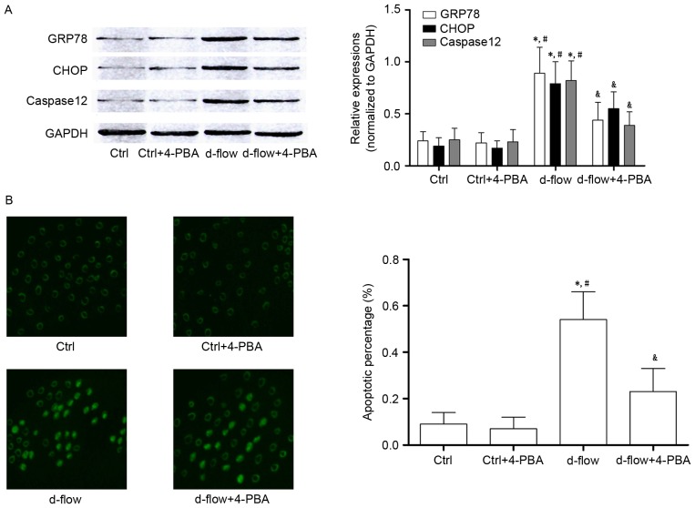
Figure 3.
Analysis of the effects of treatment with 4-PBA in HAECs exposed to d-flow. (A) Results of western blotting of GRP78, CHOP, caspase 12 and GAPDH in HAECs treated with 4-PBA and/or d-flow. The relative expression of GRP78 (white columns) and CHOP (black columns) in HAECs treated with 4-PBA and/or d-flow was normalized to GAPDH. (B) The left panel exhibits the captured images from the terminal deoxynucleotidyl transferase dUTP nick end labeling assay of HAECs treated with 4-PBA and/or d-flow. The detected apoptotic percentages of HAECs treated with 4-PBA and/or d-flow were quantified. Magnification, ×200. *P<0.05 vs. Ctrl; #P<0.05 vs. Ctrl+4-PBA; &P<0.05 vs. d-flow. HAECs, human aortic endothelial cells; d-flow, disturbed blood flow; GRP78, 78 kDa glucose-regulated protein; CHOP, DNA damage-inducible transcript 3 protein; 4-PBA, 4-phenylbutyric acid; Ctrl, control.

