Figure 3.
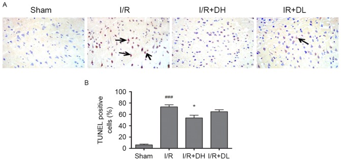
Effect of DhHP-6 on neuronal apoptosis of the ischemic cortex in rats 22 h post-reperfusion. (A) TUNEL staining was used to identify apoptotic cells in the parietal cortex. Arrows indicate TUNEL-positive cells (magnification, ×200). (B) As demonstrated in the bar graphs, the apoptotic index indicates the percentage of TUNEL-positive cells in the ischemic cortex. The percentage of TUNEL-positive cells in the I/R+DH group was significantly lower, compared with that in the I/R group. Data are demonstrated as the mean ± standard deviation. One-way analysis of variance and Tukey's post hoc test were performed. ###P<0.001, vs. Sham group; *P<0.05, vs. I/R group (n=5). DhHP-6, deuterohemin His peptide-6; DH, 1 mg/kg/day DhHP-6; DL, 0.1 mg/kg/day DhHP-6.

