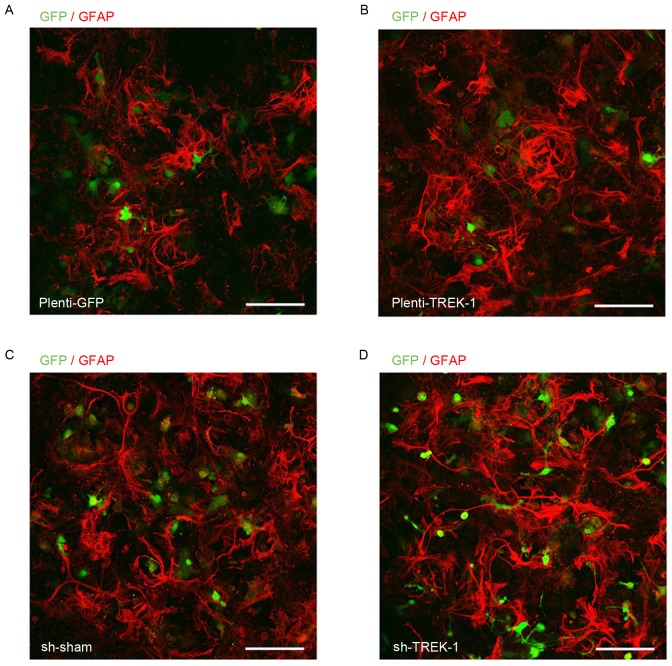
Figure 2.
Microphotographs indicating GFAP staining (red) and lentivirus infection (green) in astrocytes. Astrocytes were successfully infected with (A) Plenti-GFP lentivirus as an overexpression control, (B) Plenti-TREK-1-GFP lentivirus for TREK-1 overexpression, (C) shRNA-sham-GFP lentivirus as a knockdown control and (D) shRNA-TREK-1-GFP lentivirus for knockdown of TREK-1. Scale bar, 200 µm. GFAP, glial fibrillary acidic protein; GFP, green fluorescent protein; TREK-1, TWIK-related K+ channel; shRNA, short hairpin RNA.

