Figure 3.
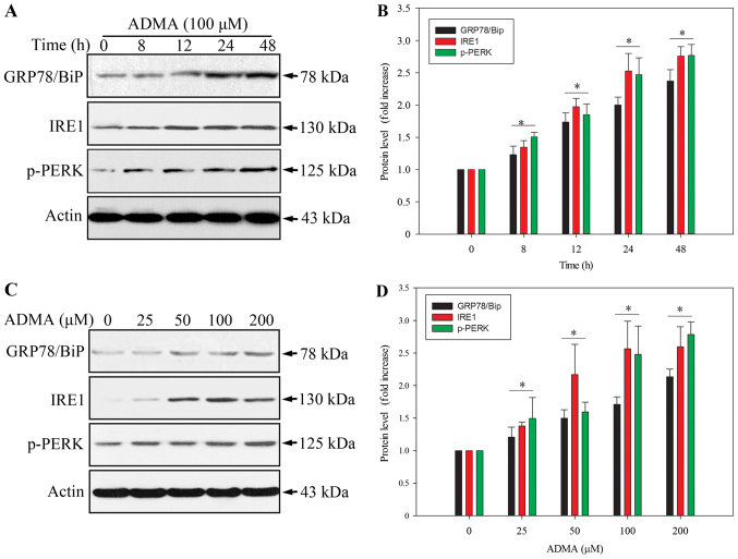
ADMA induces ER stress-associated cell death in HUVECs. Cells were incubated with 100 µM ADMA for the indicated time periods (A) or with the indicated ADMA concentration (0–200 µM) for 24 h (B). GPR78/BiP expression was analyzed by immunoblotting. (C) Cells were incubated with 100 µM ADMA for the indicated time periods. The protein levels of phosphorylated PERK and IRE1 were examined by immunoblotting. Actin was used as a loading control. (D) After preincubation with various ADMA concentrations (0–200 µM) for 24 h, proteins were extracted and analyzed for p-PERK and IRE1 by western blotting. Results are the mean ± SD (n=3). *P<0.05 compared with control.

