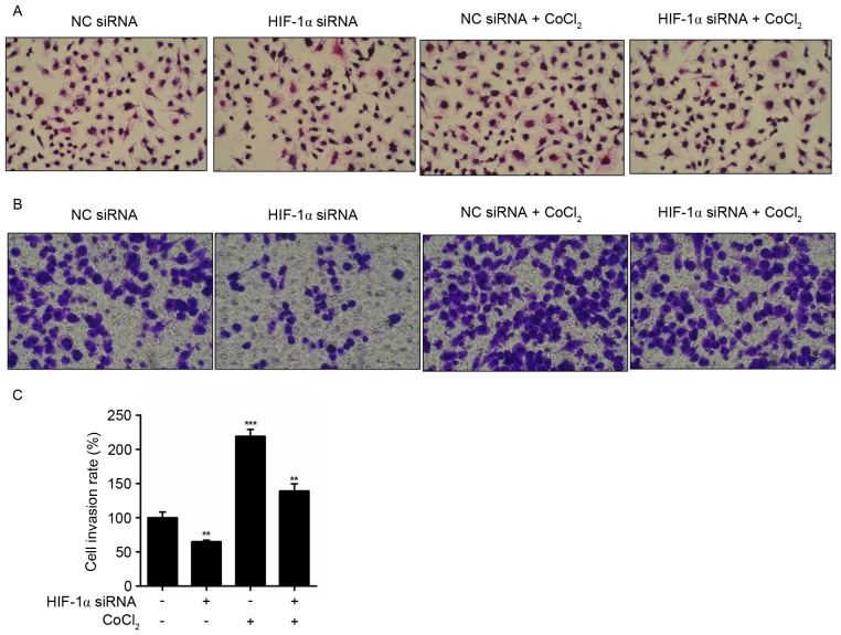
Figure 4.
Effects of HIF-1α siRNA on morphology and invasion in MiaPaCa2 cells treated with CoCl2. (A) The morphology of cells was detected by hematoxylin and eosin staining and observed from five randomly selected microscopic visual fields (magnification, ×200) following treatment with or without CoCl2 for 24 h in the presence or absence of HIF-1α siRNA. (B) Cell invasion was detected in MiaPaCa2 cells following treatment with or without CoCl2 for 24 h in the presence or absence of HIF-1α siRNA. Invaded cells were stained with crystal violet and observed from five randomly selected microscopic visual fields (magnification, ×200). (C) Number of invaded cells were counted and expressed as cell invasion rate (%) compared with those untreated cells without CoCl2 or HIF-1α siRNA. Data were representative of three independent experiments and expressed as the mean ± standard error of the mean. **P<0.01, ***P<0.001. HIF-1α, hypoxia inducible factor-1α; si, small interfering; NC, negative control; CoCl2, cobalt II chloride.

