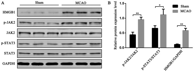
Figure 2.
Changes in HMGB1, JAK2, p-JAK2, STAT3, and p-STAT3 expression in brain tissue of rats after cerebral ischemia. Sham group and MCAO group, 3 rats in each group. The brain tissues were harvested 24 h after operation and change of HMGB1, JAK2, p-JAK2, STAT3, and p-STAT3 contents in the cerebral tissue homogenate was measured using western blotting. (A) Protein bands of cerebral tissue homogenate; (B) Relative expression of the target protein in the homogenate of brain tissue was calculated using the ImageJ image analysis software (Bethesda, MD, USA) (**P<0.01, *P<0.05).

