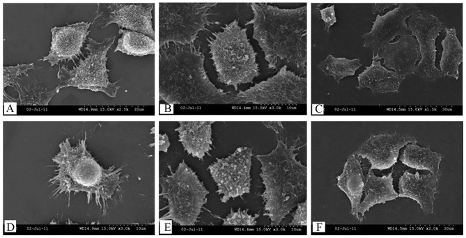
Figure 5.
Surface morphological characteristics of cells following (A and D) 0, (B and E) 24 and (C and F) 72 h of serum-starvation, detected by scanning electron microscopy. (A) Synapse connections (magnification, ×2500) and (D) a uniform distribution of microvilli on their surfaces (magnification, ×3,000) among control cells. After 24 h of serum-starvation, (B) the microvilli numbers were reduced (magnification, ×3,000) and (E) a smoothening of the cell surface was observed (magnification, ×3,000). After 72 h of serum-starvation, (C) the cell membrane showed breakage (magnification, ×1,500) and (F) the microvilli were absent (magnification, ×2,000).

