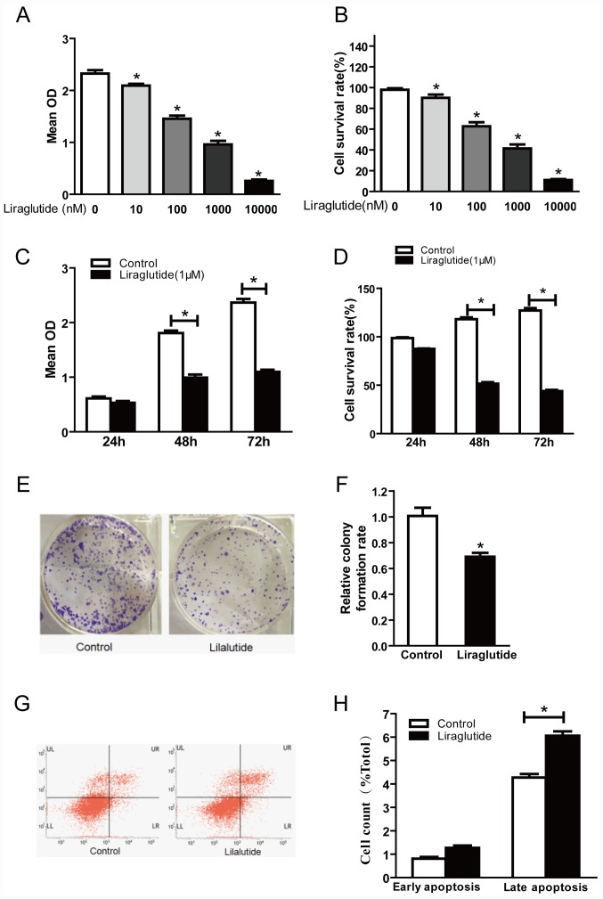
Figure 1.
Liraglutide inhibits the proliferation and promotes apoptosis in the MCF-7 breast cancer cell line. (A) CCK-8 assay results demonstrated that with increasing liraglutide concentration, the mean OD values were reduced. (B) Cell survival rates in each group were calculated following CCK-8 assays. (C) When 1,000 nM liraglutide was employed for 48 and 72 h, cell proliferation was markedly inhibited. (D) Cell survival rates in each group were calculated following CCK-8 assays. (E) Representative images of cell colony formation assays. (F) Quantification of cell colony formation assay results confirmed that the colony formation capacity decreased significantly following treatment with 1,000 nM liraglutide. (G) Representative flow cytometry plots in control cells or cells treated with 1,000 nM liraglutide. LR quadrant represents early apoptotic cells, and UR quadrant represents late apoptotic cells. (H) Quantified flow cytometry results demonstrated that the percentage of late apoptotic cells in the liraglutide treatment group increased significantly compared with the control group. Data are presented as the mean ± standard deviation. For parts A and B, *P<0.05 vs. 0 nM liraglutide group; for parts C, D, F and H, *P<0.05, as indicated by brackets. CCK-8, Cell Counting Kit-8; OD, optical density.

