Figure 2.

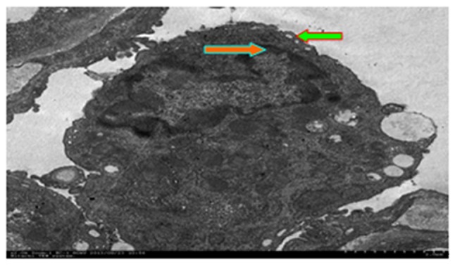
Apoptotic morphology detected using a transmission electron microscope (magnification, ×8,000). The red arrow indicates chromatin margination and the green arrow indicates condensation. The results show microvilli disappearance.

Apoptotic morphology detected using a transmission electron microscope (magnification, ×8,000). The red arrow indicates chromatin margination and the green arrow indicates condensation. The results show microvilli disappearance.