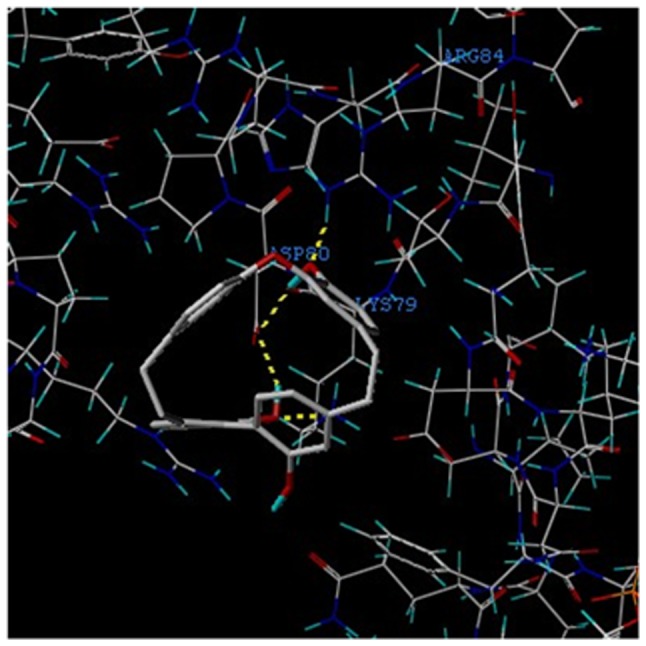
Figure 4.

Theoretical binding mode of Riccardin D to the NF-κB-p65 protein. Superimposition of the native crystal structure of Riccardin D (grey) and the best docked conformation obtained by AutoDock Vina software (red). Representation of the 2 competitive binding sites, ASP-80 and LYS-79 (blue), between Riccardin D and NF-κB-p65 as obtained from AutoDock analysis. The dashed yellow lines represent the hydrogen bonds of the compound with residues of surrounding amino acids. NF-κB, nuclear factor-κB; p-, phosphorylated.
