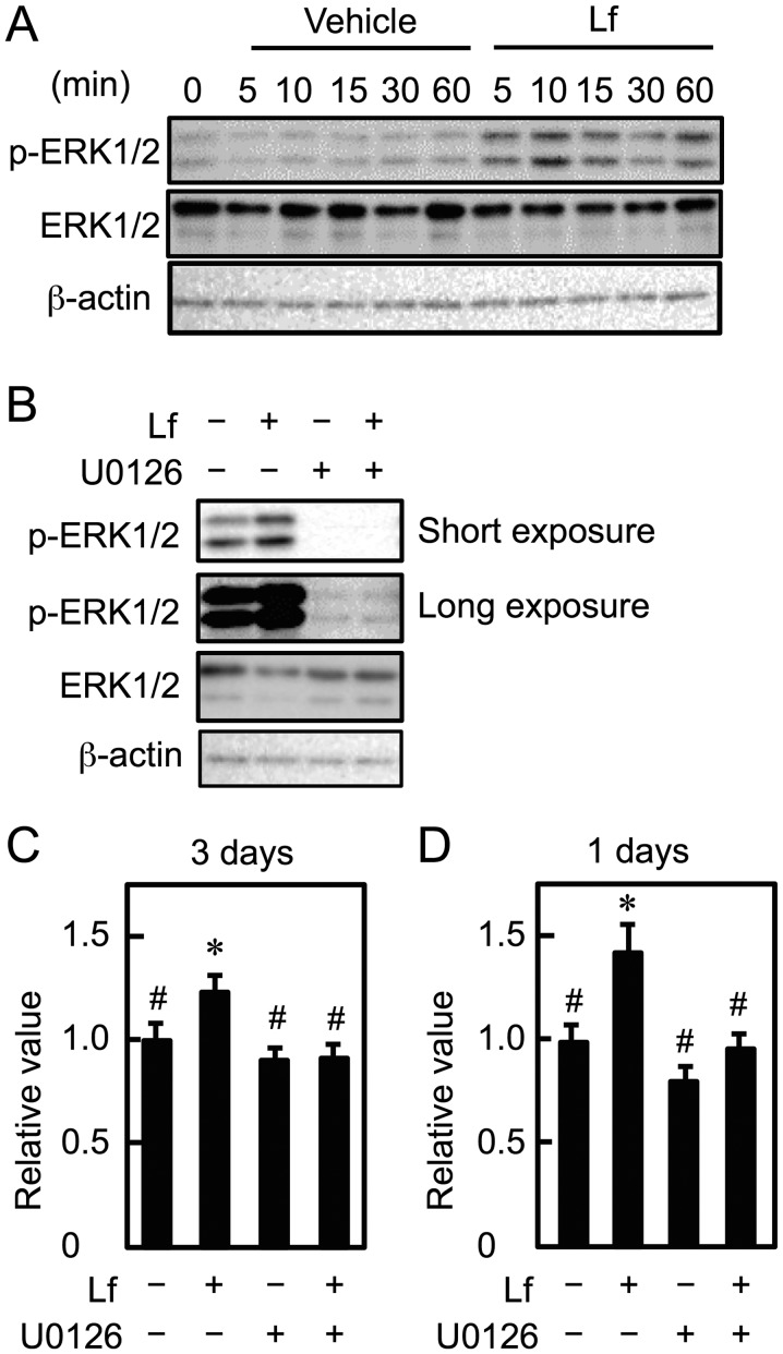
Figure 2.
Involvement of ERK1/2 in the Lf-induced proliferation of myoblasts. (A) Myoblasts were incubated with Lf (100 µg/ml) for the indicated times. The expression of ERK1/2 and phosphorylated ERK1/2 (p-ERK1/2) was analyzed by western blots with anti-ERK1/2 and anti-p-ERK1/2 antibodies. The expression of β-actin was analyzed as a loading control. (B) Myoblasts were incubated with Lf in the presence of U0126 for 10 min. The expression of ERK1/2 and p-ERK1/2 was analyzed by western blots. The expression of β-actin was analyzed as a loading control. The data for p-ERK1/2 expression level were obtained as digitized images for short (upper panel) or long (middle panel) exposure times. (C) Myoblasts were cultured with Lf (10 µg/ml) in the presence of U0126 for three days. (D) Myoblasts were cultured with U0126 in the presence of Lf (10 µg/ml) for the first day and in the absence of Lf for the next two days. (C and D) Cell viability was determined by the alamarBlue assay. Data are expressed as relative values (FI of the experimental group divided by FI of the vehicle group (-Lf, -U0126). Values are indicated as the mean ± standard deviation (n=4). Statistically significant differences were determined by one-way ANOVA and Tukey's post-hoc test. *P<0.001 vs. vehicle group (-Lf, -U0126). #P<0.001 vs. Lf group (+Lf, -U0126). Each result is representative of three independent experiments (C) or two independent experiments (D). ERK, extracellular signal-regulated kinase; FI, fluorescence intensity; Lf, lactoferrin; p-, phosphorylated.

