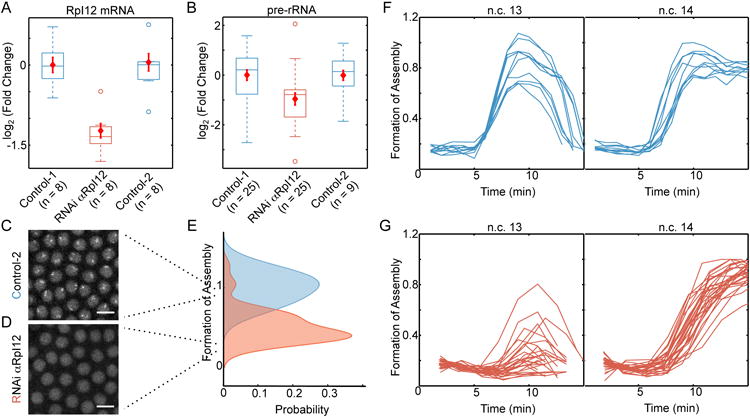
Figure 7. Reduction in rRNA transcription delays nucleolus formation.

A. Injection of wild-type embryos with RNAi against RpI12 reduces the amount of this transcript to approximately 45% of that of un-injected embryos (Control-1) or embryos injected with RNAi against RpI135 (Control-2). B. The amount of pre-rRNA is determined by RT-qPCR on 25 individual control-1 embryos, 25 embryos injected with RNAi αRpI12, and nine of Control-2 embryos at 10 min into n.c. 13. The amount of pre-rRNA is reduced by nearly 2 fold in the embryos injected with RNAi for RpI12 compared to the two controls (P < 0.005 by Student's t-test). Red diamonds in A-B show mean ± SEM for each group. Representative images of n.c. 13 for a Control-2 embryo with normal nucleolus formation (C) and RpI12 RNAi embryo with delayed nucleolus formation (D). Scale bars, 10 μm. E. Formation of assemblies was measured for 39 individual RpI12 RNAi embryos (orange) and 39 un-injected embryos (blue) at 10 min into n.c. 13. Images shown in C and D score 0.9 and 0.3 for Formation of Assembly. F. Time evolution of formation of fibrillarin assemblies in the wild-type embryos (n=10, reproduced from Figure 4F) and G. RpI12 RNAi embryos (n=28) during n.c. 13-14. Each line depicts an individual embryo. Time zero marks the end of mitosis.
