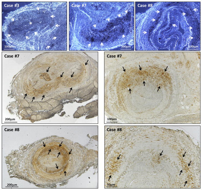
FIGURE 2.
Varicella-zoster virus (VZV) positivity in temporal artery biopsies. Examples of immunohistochemistry-positive results from 3 giant cell arteritis (GCA)-positive patients. The top row includes samples examined by wet mount; the center and bottom rows contain samples examined by dry mount at 2 magnifications. Zoster antigen was detected with monoclonal antibody 3B3 against major zoster protein E (gE) and appears as darker stained areas indicated by white arrows in the top row (wet mounts) and by black arrows in the center and bottom rows (dry mounts). Case #3 shows VZV-positive staining located in a branch artery off the main vessel (asterisk). Case #7 (center row) shows VZV-positive staining located mainly in the medial layer. Case #8 shows VZV-positive staining located in the intimal, medial and adventitial layers. Scale bars are shown in each image.

