. 2018 Mar 16;7:e33680. doi: 10.7554/eLife.33680

Figure 1. notum RNAi shifts the site of eye regeneration anteriorly.

(A) Animals were treated with notum or control dsRNA every 2–3 days for (top) 40 days in the absence of injury or (bottom) for four times over 9 days followed by decapitation and 28 days of head regeneration as indicated. notum(RNAi) animals produced an anterior set of eyes (129/143 notum(RNAi) homeostasis animals and 187/200 notum(RNAi) regenerating head fragments, yellow arrowheads) and retained a pre-existing set of eyes (white arrowheads). (B) FISH to detect expression of opsin and tyrosinase. (C) anti-ARRESTIN immunostaining to detect photoreceptor neuron axons. (D) Surgical removal of eyes in control and notum(RNAi) animals generated by homeostatic RNAi treatment as in (A), showing individuals at 1 day after surgery to confirm successful removal (white arrowheads) and 14 days to assess regeneration. In notum(RNAi) animals, 40/40 anterior supernumerary eyes regenerated after removal (yellow arrowheads) and 37/38 posterior pre-existing eyes failed to regenerate (red arrowheads). Right, FISH of ovo confirms lack of eye cells produced in the region of the resected notum(RNAi) posterior eyes. Scale bars, 300 microns.

Figure 1.

Figure 1—figure supplement 1. Regenerative and non-regenerative eyes both mediate negative phototaxis.

Figure 1—figure supplement 1.

(A) Phototaxis behavior was measured by measuring the time of transit across an arena illuminated from one side. (B) Illustration of outcomes in the assay. (C) Control and notum(RNAi) animals were examined in phototaxis assays after no treatment, removal of all eyes or removal of only either the supernumerary or pre-existing eyes. (D) Time of transit data for animals after surgeries. Only removal of all eyes in either control or notum(RNAi) animals resulted in lack of negative phototaxis. (E) Quantification of data in D showing average time from the timeseries spent in the blue quadrant (greater than 100 mm from the illuminated side). **p<0.01 by 2-tailed t-test; n.s. denotes p>0.05 from the same test.

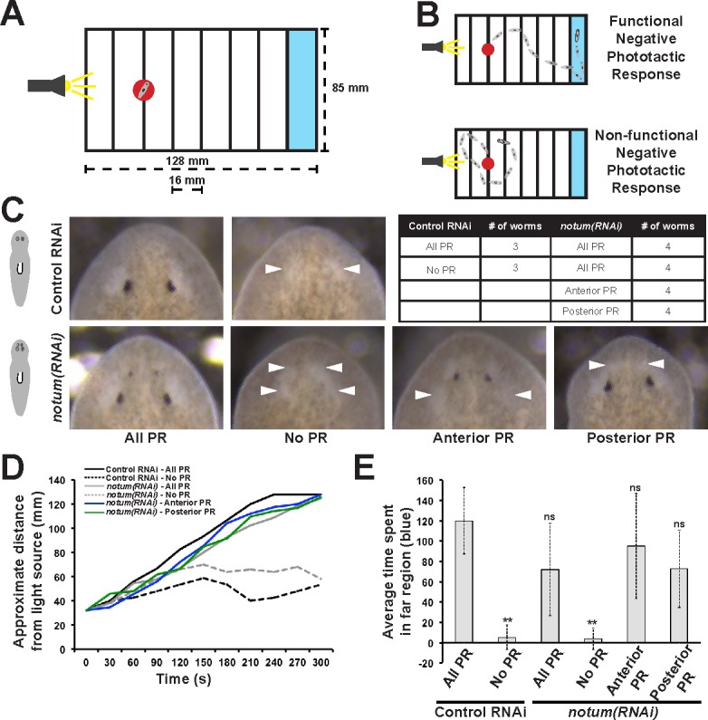
Figure 1—figure supplement 2. Additional controls for structure and regenerative ability of eyes from notum(RNAi) animals.

Figure 1—figure supplement 2.

(A) Homeostasis notum(RNAi) animals were generated by dsRNA feeding for 40 days followed by surgeries as indicated by cartoons. Removal of both a supernumerary anterior eye and a posterior pre-existing eye resulted in regeneration only of an eye at the anterior eye position. Likewise, removal of all four eyes from such animals resulted in eye regeneration at the anterior position. (B) 4-eyed notum(RNAi) animals were generated by allowing decapitated head fragments to regenerate for 28 days, then tested for eye regeneration behavior. In such animals, the pre-existing eyes fail to regenerate whereas supernumerary eyes have regenerative ability.

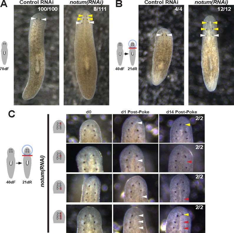
Figure 1—figure supplement 3. Prolonged notum RNAi and surgical strategies can create additional sets of ectopic eyes that track with regenerative ability.

Figure 1—figure supplement 3.

(A) At a low frequency, notum(RNAi) homeostasis animals form an additional set of ectopic eyes (8/111) animals at 70d RNAi to generate 6-eyed animals. (B) Alternatively, 6-eyed animals can be produced at higher frequency by decapitating 4-eyed notum(RNAi) animals generated by homeostatic inhibition. (C) Experiments to test regenerative ability of the three sets of eyes from 6-eyed notum(RNAi) animals. Only the anterior-most set of eyes can regenerate (top panels vs. middle panels). Removal of a set of three eyes from the same side of these worms results in regeneration only of the anterior-most set of eyes.