. 2018 Mar 16;7:e33680. doi: 10.7554/eLife.33680

Figure 5. Modulation of other patterning factors alters the sites of eye or pharynx regeneration.

(A) Simultaneous inhibition of wnt11-6 and fzd5/8-4 resulted in the formation of ectopic eyes posterior to the original eyes. Removal of the supernumerary, posterior eyes resulted in regeneration (7/10 animals) whereas removal of the original, anterior eyes did not result in regeneration (11/11 animals), p=0.001 by Fisher’s exact test. (B) wntP-2(RNAi);ptk7(RNAi) animals form a supernumerary posterior pharynx while retaining a pre-existing central pharynx. Cartoons denote amputations used to test regenerative ability of pre-existing or supernumerary pharynges from control or wntP-2(RNAi);ptk7(RNAi) animals. wntP-2(RNAi);ptk7(RNAi) animals were prepared by dsRNA feeding for 3 weeks, then amputated using repeated punctures centrally in a box shape around the target pharynx. Regeneration of the wntP-2(RNAi);ptk7(RNAi) supernumerary posterior pharynx occurred at frequencies close to those of control animal pharynges, but regeneration ability of the wntP-2(RNAi);ptk7(RNAi) pre-existing anterior pharynx was markedly reduced (p=0.03 by Fisher’s exact test).

Figure 5.

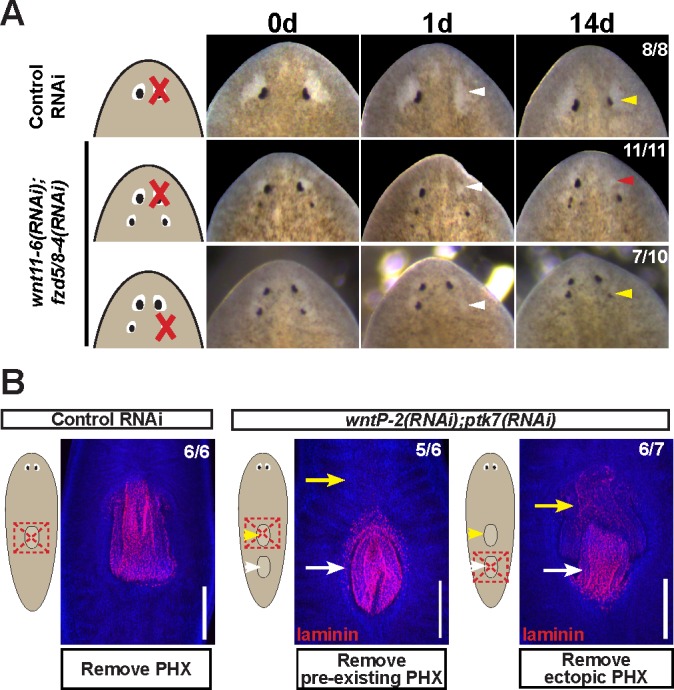
Figure 5—figure supplement 1. Additional staining and verification of the ectopic posterior eye phenotype of wnt11-6(RNAi);fzd5/8-RNAi(RNAi) animals.

Figure 5—figure supplement 1.

(A) Live images of wnt11-6(RNAi);fzd5/8-RNAi(RNAi) animals after 35 days of RNAi feeding. (B) Images of control and wnt11-6(RNAi);fzd5/8-RNAi(RNAi) animals staining for ARRESTIN protein and eye53-1 and eye53-2 probes.

Figure 5—figure supplement 2. Tests to determine the homeostatic potential of supernumerary eyes and pharynges formed by RNAi of Wnt pathway components.

Figure 5—figure supplement 2.

(A) wnt11-6(RNAi);fzd5/8-4(RNAi) animals with ectopic eyes were generated by dsRNA feeding for 40 days and animals were tracked for a subsequent 100 days after feeding. 3/4 animals maintained two sets of eyes during this time and 1/4 animals maintained three eyes during this time. (B) Four-eyed wnt11-6(RNAi);fzd5/8-4(RNAi) animals were generated as in (B), injected with BrdU then fixed and stained 7 days later with opsin and tyrosinase riboprobes and anti-BrdU antibody. notum(RNAi) animals labeled with BrdU had BrdU +cells in both the supernumerary posterior and pre-existing anterior eyes (5/5 animals), similar to control individuals (5/5 animals). (C) Tests using BrdU to determine homeostatic maintenance ability of new and pre-existing pharynx in wntP-2(RNAi);ptk7(RNAi) animals prepared as in Figure 5C then pulsed with BrdU prior to fixing and staining 7 days later with anti-BrdU antibody and laminin riboprobe that labels pharyngeal tissue. Both pharynges acquired BrdU +cells during the pulse (9/9 animals). Bars, 100 microns.

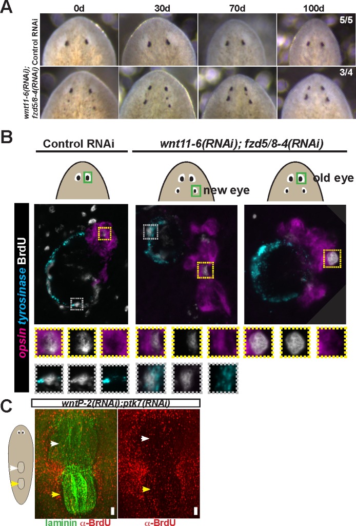
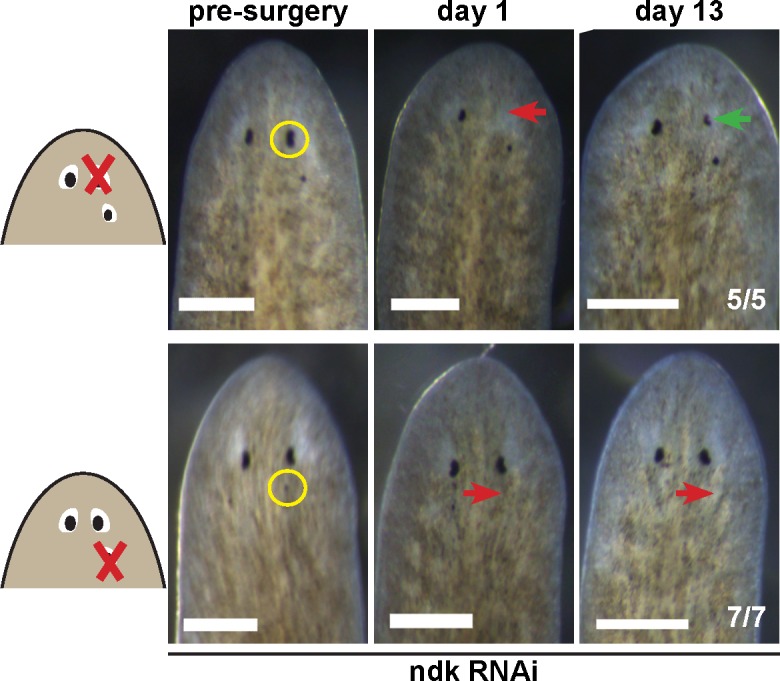
Figure 5—figure supplement 3. Tests to determine the regenerative potential of eyes in ndk(RNAi) animals.

Figure 5—figure supplement 3.

Animals were fed ndk dsRNA 6 times over 2 weeks then decapitated and regenerating head fragments scored 21 days later for ectopic eyes (15/31 animals). Animals displaying this phenotype were selected for eye resection to remove either an original anterior eye or a supernumerary posterior eye. Removal of the anterior eye resulted in regeneration (5/5 animals), while regeneration was not observed after removal of posterior eyes (7/7) as scored 14 and 21 days later.