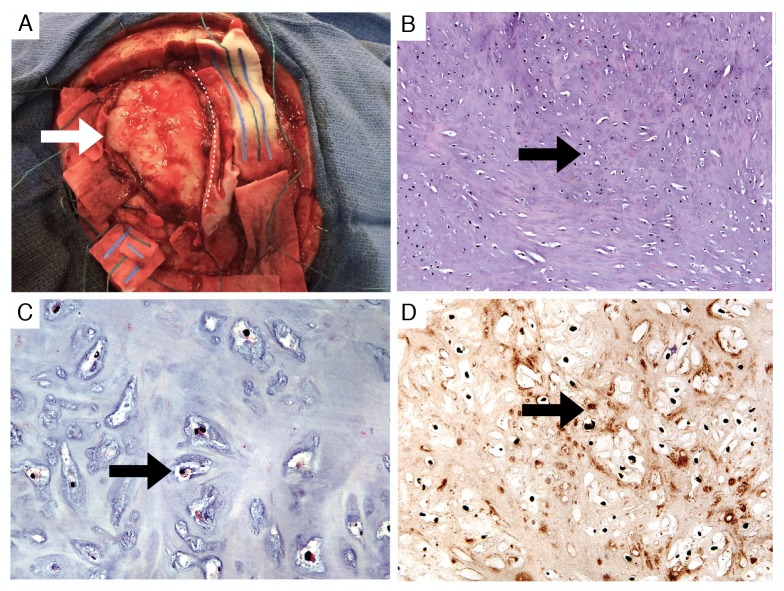
Figure 3. Gross appearance and histology of the tumor.
Appearance of the mass (white arrow) during piecemeal resection (A). The superior sagittal sinus is protected under a midline patty (dotted line). Routine histologic sections demonstrated cytologically bland chondrocytes (B) 100x and (C) 400x H&E (black arrows). Immunohistochemistry for S100 protein was positive, confirming a chondrocyte origin (D) 200x, (black arrow).
H&E: hematoxylin and eosin

