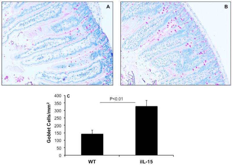
Figure 4. Induced Goblet cells hyperplasia in the jejunum of iIL-15 mice.

PAS stained jejunum tissue section of WT (A) and iIL-15 (B) mice detected Goblet cells (Photomicrograph presented are ×400 of original magnification). Morphometric quantitation of Goblet cells in iIL-15 mice compared to wild type mice (C). The data was expressed as mean ± SD; Experiment “n”= 3, 4 mice/group.
