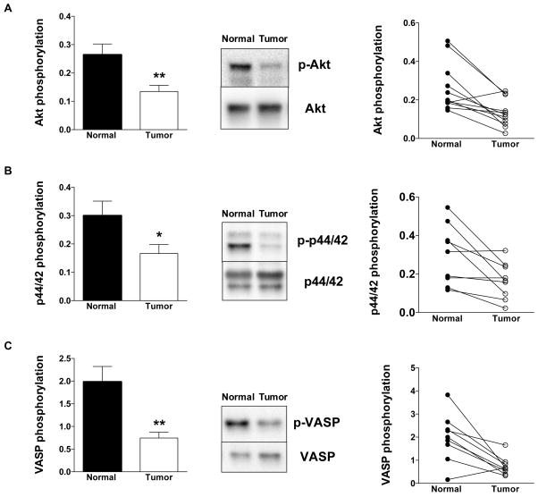
Figure 11. Expression of (A) pAkt and Akt, (B) p-p44/42 MAPK and p44/42 MAPK, and (C) pVASP and VASP in normal surrounding tissue (Normal) and human colon cancer biopsies (Tumor).
For each enzyme, representative Western blots, summary of expression data (mean±SEM; phosphorylated forms normalized to non-phosphorylated) from all samples analyzed (where detectable signal was found) and individual paired sample analysis is shown. *P<0.05 Tumor vs. Normal; n=8.

