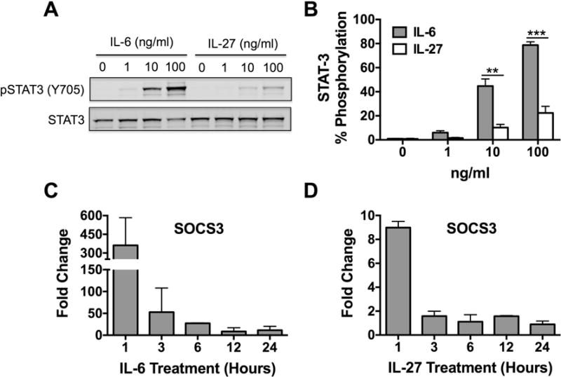
Figure 2. IL-6 induces STAT3 phosphorylation and SOCS3 expression via classical signaling in human skin mast cells.

To determine the functionality of membrane-bound gp130 and IL-6Rα, STAT3 phosphorylation and SOCS3 expression was analyzed in IL-6-treated human skin mast cells. For comparison, parallel studies were performed with IL-27, which also utilizes gp130. STAT3 phosphorylation was determined by SDS-PAGE and Western blotting of whole cell lysates prepared from skin mast cells treated with IL-6 or IL-27 at 1, 10, or 100 ng/ml for 15 min (A). STAT3 phosphorylation was quantified with Western blot band intensities (B). SOCS3 expression was determined by quantitative real-time PCR of total RNA from skin mast cells treated with IL-6 (100 ng/ml) (C) or IL-27 (D) for 1, 3, 6, 12, or 24 h. Fold change was calculated using the 2ΔΔCt method with B2M as the reference gene. >2-fold change was considered significant. Bars represent mean ± S.E.M. of values from 3 separate experiments with mast cells from different donors. Significance was determined with Student’s t-test. **, p<0.01 and ***, p<0.001. Representative blot is shown.
