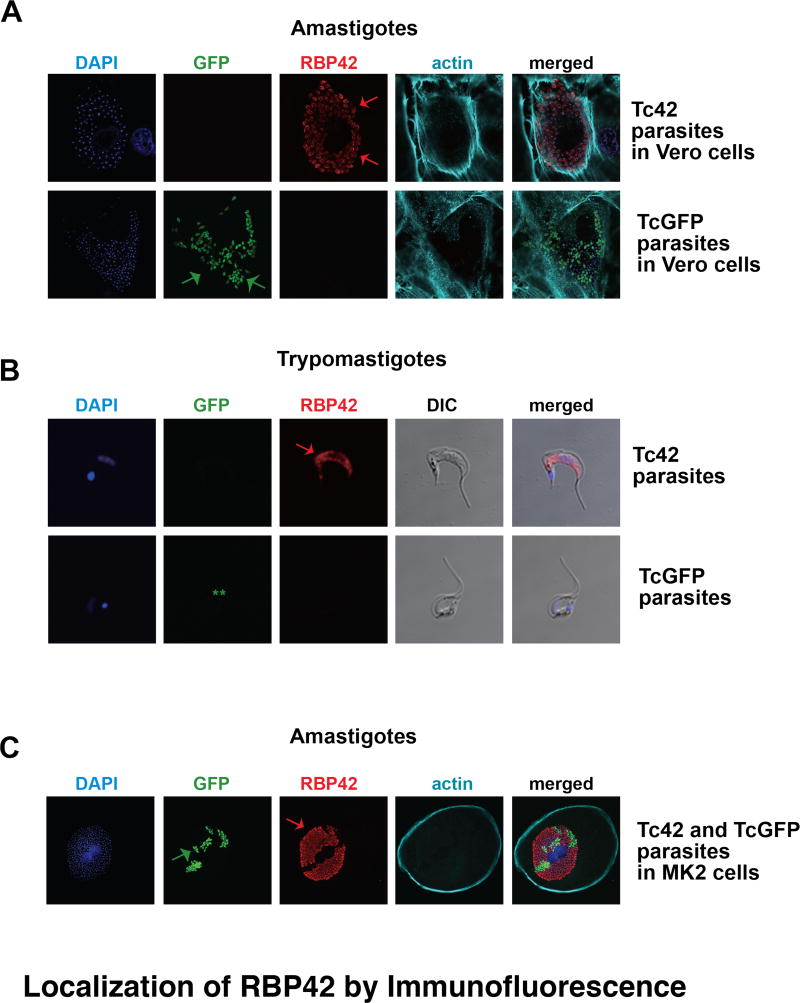
Figure 5. Immunolocalization of RBP42 in amastigote and trypomastigote stages of the parasite life cycle.
Panel A and B: Immunolocalization of RBP42 in amastigote and trypomastigote stages of the parasite life cycle. Staining is as described in Figure 4, with the addition of phalloidin staining to visualize host cell periphery. In Panel B, the two asterisks (**) indicate that the GFP signal was lost in these parasites. Arrows indicate staining of RBP42 and GFP fluorescence.
Panel C: LLC-MCK cells were co-infected with similar numbers of Tc42 and TcGFP trypomastigotes, which then differentiated into amastigotes. Staining is as described in Panel A and B.

