Figure 1.

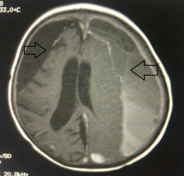
Head MRI with contrast showing bilateral subdural empyema in the frontotemporoparietooccipital region (arrows), slight parenchymal hemorrhage, communicating hydrocephalus and meningoencephalitis

Head MRI with contrast showing bilateral subdural empyema in the frontotemporoparietooccipital region (arrows), slight parenchymal hemorrhage, communicating hydrocephalus and meningoencephalitis