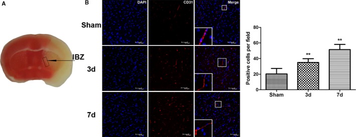
Figure 1.

Neovascularization around ischemic foci after cerebral ischemia/reperfusion in mice. A, Schematic drawing of ischemic foci and the infarction border zone (IBZ) after cerebral ischemia/reperfusion in mice. B, Compared with the sham‐operated control group, the number of CD31‐stained vascular endothelial cells around the ischemic foci increased significantly 3 and 7 days after middle cerebral artery occlusion/reperfusion in the treated mouse group; this number gradually increased with time after ischemia/reperfusion (n=6, **P<0.01). d indicates days; DAPI, 4′,6‐diamidino‐2‐phenylindole.
