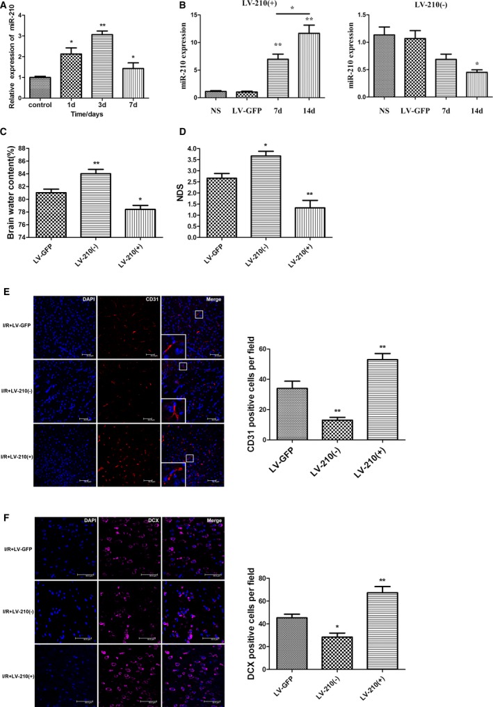
Figure 4.

The expression of miR‐210 was significantly increased after the cerebral ischemia/reperfusion (I/R) in mice, and miR‐210 overexpression promoted neovascularization and neural precursor cell migration toward ischemic foci to alleviate neural function damage. A, The expression of miR‐210 in the ischemic brain tissue of the middle cerebral artery occlusion (MCAO) group was quantified using quantitative real‐time–polymerase chain reaction (qRT‐PCR) at 1, 3 and 7 days after I/R. Compared with the control group, miR‐210 expression levels were significantly higher in the MCAO group, with the highest level being observed at day 3 (*P<0.05, **P<0.01). B, Lentivirus (3 μL) with the miR‐210 overexpression construct (LV‐210[+]) or a miR‐210 inhibitor construct (LV‐210[−]) was injected into the right striatum of the mice. An empty vector (LV‐GFP) or normal saline (NS) injection was used as a control. The expression of miR‐210 in the right striatum was detected by qRT‐PCR on days 7 and 14 after the injection. The expression of miR‐210 was increased significantly on days 7 and 14 after LV‐miR‐210 injection (**P<0.01). The expression of miR‐210 was significantly higher on day 14 than on day 7 (*P<0.05). The expression on miR‐210 was significantly decreased on day 14 after injection of the LV‐miR‐210 inhibitor (*P<0.05). C and D, The right striatum of the mice was injected using a stereotactic apparatus with 3 μL of lentivirus with LV‐210(+), LV‐210(−), or an empty vector (LV‐GFP). These mice were used to establish the I/R model 14 days after virus injection. The neurological deficit score (NDS) was obtained 3 days after I/R, and the brain water content (BWC) on the ischemic side of the brain was calculated. Compared with the control group injected with LV‐GFP, the overexpression of miR‐210 attenuated I/R‐induced cerebral edema, whereas the BWC was higher in the group with miR‐210 expression inhibition. The NDS was significantly decreased in the miR‐210 overexpression group and increased in the miR‐210 inhibition group (n=6, *P<0.05, **P<0.01). E and F, The right striatum of the mice was injected using a stereotactic apparatus with 3 μL of LV‐210(+) or LV‐210(−), or LV‐GFP, and the mice were used to establish the I/R model at 14 days after virus injection. At 7 days after I/R, the vascular endothelial cell‐ and DCX (doublecortin)–positive neuroblast numbers around the ischemic foci increased significantly in the miR‐210 overexpression group compared with the control group injected with LV‐GFP, whereas these numbers decreased significantly in the miR‐210 inhibition group (n=6, *P<0.05, **P<0.01). d indicates days; DAPI, 4′,6‐diamidino‐2‐phenylindole.
