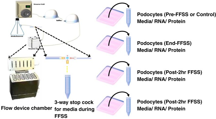
Fig. 2.
Left: outline of the apparatus used for applying fluid flow shear stress (FFSS) to podocytes. The “fluid chamber” contains cells grown on six glass slides placed for FFSS studies, and a three-way stopcock is attached to collect media during application of FFSS. Right: experiment design for collecting supernatant culture medium, cells for RNA, and protein extraction at four different experimental time points (Pre-FFSS or Control, End-FFSS, Post-2 h FFSS, and Post-24 h FFSS).

