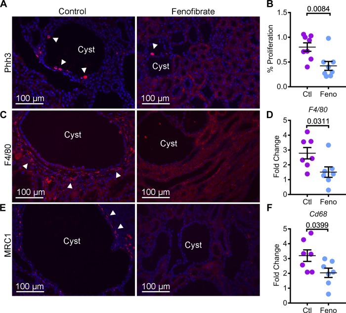
Fig. 5.
Fenofibrate treatment inhibits cyst proliferation and interstitial inflammation. A and B: kidney sections from control and fenofibrate-treated Pkd1RC/RC mice were stained with phospho-histone H3 (Phh3), a marker of proliferation. Immunofluorescence analysis and quantification of Phh3-positive cells (red, arrowheads) showed that proliferation was reduced in the kidneys of fenofibrate-treated (n = 8) compared to control-treated mice (n = 8). To assess inflammation, kidney sections from control and fenofibrate-treated Pkd1RC/RC mice were stained using an antibody against F4/80, a pan-macrophage marker, and MRC1, a marker of M2-like macrophages. C: immunofluorescence analysis revealed that the abundance of F4/80-positive inflammatory cells (red, arrowheads) was reduced in fenofibrate-treated Pkd1RC/RC mice compared to control-treated Pkd1RC/RC mice. D: Q-PCR analysis also demonstrated decreased F4/80 expression in fenofibrate-treated Pkd1RC/RC mice (n = 7) compared with control-treated Pkd1RC/RC mice (n = 7). E: expression of MRC1 (red, arrowheads) was reduced in fenofibrate-treated Pkd1RC/RC mice. F: Q-PCR analysis demonstrated that the expression of Cd68, another marker of macrophages, was downregulated in fenofibrate-treated (n = 8) compared with control-treated (n = 7) Pkd1RC/RC mice. Ctl, control; feno, fenofibrate. Error bars indicate means ± SE.

