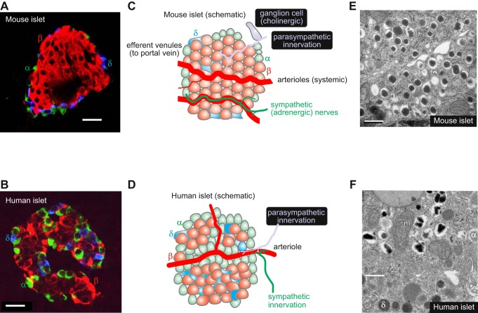
FIGURE 1.
A and B: immunohistochemistry of mouse (A) and human (B) pancreatic islets (red, insulin; green, glucagon; blue, somatostatin). Images provided by Dr A Clark, Oxford. Scale bars: 20 μm. C and D: schematic of mouse (C) and human (D) islets highlighting differences in blood supply, innervation, and islet cell distribution. The α- (green), β- (red), and δ-cells (blue) are indicated. Also illustrated (C, gray) is a pancreatic ganglion cell (613). E and F: electron micrographs of mouse (E) and human (F) β-cells. Scale bars: 500 nm. In F, the β-cell is surrounded by a δ- and an α-cell (granules indicated by α and δ). Electron micrographs provided by Prof. L. Eliasson, Lund (E), and Dr. A. Clark, Oxford (F). m, Mitochondrion; l, lipfuscin body; sg, secretory granule.

