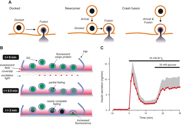
FIGURE 23.
A: different modes of insulin granule fusion. Release of previously ‟dockedˮ granules upon stimulation (left), release of granules not originally docked with the plasma membrane but that are recruited to the plasma membrane and undergo exocytosis following a variable period of being docked (‟newcomer,ˮ middle) and granules that arrive at the plasma membrane and are instantly released without being docked (‟crash fusion,ˮ right). [Modified from Shibasaki et al. (617).] B: imaging of secretory granules by TIRF microscopy. Red area indicates the evanescent field. Illumination leads to time-dependent bleaching of fluorescently labeled secretory granules (bright green → dark green). Granules that were initially bright (at t = 0) might thus appear to vanish within a few minutes (t = 2 min) because of loss of fluorescence. When such granules subsequently undergo exocytosis, the dramatic increase in fluorescence as the fluorophore moves further into the evanescent field will result in the reappearance of fluorescence giving the impression that granules released by glucose (at t = 2 min) were not docked with the plasma membrane. With agents that act more quickly (like K+), exocytosis will occur (at t = 0.5 min) before granules have faded enough to make them invisible. C: insulin secretion elicited by sequential stimulation with high extracellular K+ ([K+]o) and glucose. Note that high [K+]o transiently stimulates insulin secretion and that release rates subsequently decline back towards baseline values and that subsequent addition of glucose leads to a slowly developing increase in insulin release without any sign of a 1st phase (compare FIGURE 21A). Values are means (red circles) + SE (gray shaded area). Experiment performed by M. Söderström, Oxford.

