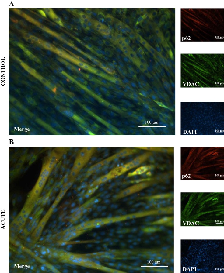
Fig. 4.
Mitophagy signaling following acute contractile activity and recovery. Fixed-cell immunofluorescence microscopy of C2C12 myotubes costained for 4′,6-diamidino-2-phenylindole (DAPI), voltage-dependent anion channel (VDAC), and nucleoporin p62 (p62) (×20 magnification). Fully differentiated myotubes were acutely stimulated for 5 h or not simulated (control) cells. Following 5 h of stimulation, there was an increase in yellow colocalization of p62 and VDAC (merge).

