
Fig. 6.
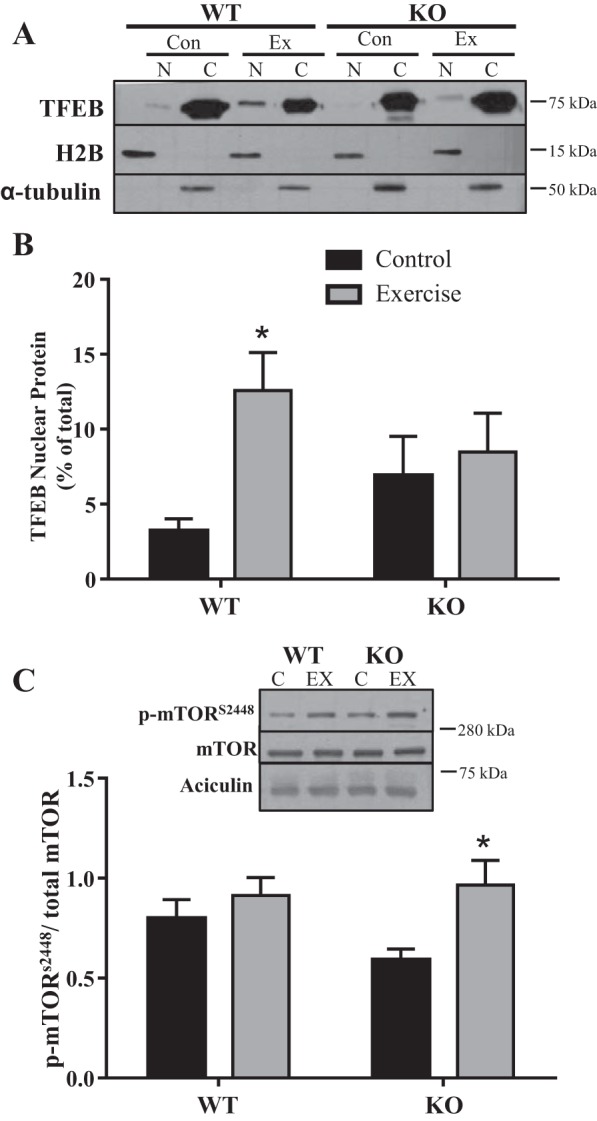
Transcription factor EB (TFEB) localization and activation. TFEB translocation into the nucleus was measured in wild-type (WT) and peroxisome proliferator-activated receptor-γ coactivator-1α (PGC-1α) knockout (KO) mice following acute exercise. Typical blot (A) and quantification of TFEB expressed as a percentage of total [cytosolic (C) and nuclear (N)] signals (B). Histone H2B and α-tubulin were used as nuclear and cytosolic purity controls (n = 7–9; *P < 0.05 WT control vs. WT exercise, 2-way ANOVA). C: mammalian/mechanistic target of rapamycin (mTOR) activation was measured by Ser2448 phosphorylation (p). Aciculin was used as a loading control (n = 7–9; *P < 0.05 KO control vs. KO exercise; 2-way ANOVA). C, control; EX, exercise.