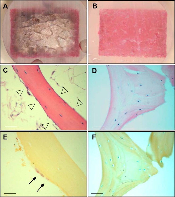
Fig. 1.
The effect of bone marrow on perfusion. Bone cores with and without bone marrow (n = 3/group) were seated in individual chambers and continuously perfused with media including Reactive red 120 for 24 h. Sagittal sections were obtained through the center of the bone cores with bone marrow (A) and without bone marrow (B). Specimens with and without bone marrow were fixed in 10% formaldehyde and decalcified with phosphate-buffered saline (pH 8.0) including 1% EDTA and 0.5% PFA, and cut into 5-µm-thick sections. Samples stained by H&E and TRAP with bone marrow (C and E) and without bone marrow (D and F). Arrowheads indicate osteoblasts and lining cells. Arrows indicate TRAP-positive osteoclasts. Scale bar, 200 µm.

